
Action Physical Therapy & Rehabilitation Inc.


GET "BACK IN ACTION"

Physical Therapist's Guide to Cuboid Syndrome

02-18-19 • April 8, 2020

Physical Therapist's Guide to Cuboid Syndrome


Cuboid syndrome is a condition caused by a problem with the cuboid bone, producing pain on the outer side, and possibly underside, of the foot. The cuboid bone is part of the calcaneocuboid joint that helps you maintain foot mobility when walking. Problems can develop with the cuboid bone that make it shift and dislodge from its normal position, causing pain and difficulty standing or walking. Cuboid syndrome most often affects athletes and dancers, although anyone can experience it. Age does not seem to play a role in developing the syndrome.

The prevalence of cuboid injuries in the United States is not clear; however, it has been diagnosed in 6.7% of patients with inversion ankle sprains (when the ankle rolls outward and the foot rolls inward). Approximately 4% of athletes who report foot injuries have symptoms from the cuboid bone. Cuboid syndrome is found in about 17% of professional ballet dancers. It can occur from trauma or without any recalled injury. Physical therapists design individualized treatment programs to help people with cuboid syndrome reduce their pain, regain lost strength and movement, and get back to their normal lives.


What Is Cuboid Syndrome?

The cuboid bone is one of the 26 bones of the foot. It is located on the outer side of the foot, about halfway between the pinky toe and the heel bone. The cuboid bone moves and shifts to a small degree during normal foot motion. Certain forceful movements or prolonged positions can cause the cuboid bone to move too far, which interferes with its normal position or motion. This causes immediate foot pain, which can feel worse when standing or walking on the foot.

Cuboid syndrome often occurs suddenly. It may occur with ankle sprains, as the foot rolls in, or when a person stomps the foot hard onto a hard surface, such as concrete, particularly if the person is wearing rigid or high-heeled shoes. Hard landings onto the feet from a jump, or falling from a height onto the feet can also create enough force to affect the cuboid bone’s position and cause a problem.

A cuboid bone injury can develop from maintaining prolonged foot positions, such as standing or walking in high heels, or remaining in a toe-pointed (ballet dancer’s) position, for a long time. Peroneal tendon problems, such as weakness, tendonitis, or tendinopathy, also can contribute to, or occur at the same time as, cuboid syndrome.

The majority of people who suffer cuboid syndrome have flat feet, although the condition can even occur in people with very high arches.


How Does it Feel?

Cuboid syndrome causes sharp pain on the outer side, and possibly underside, of the foot. The pain does not usually spread to the rest of the foot or leg. It often starts quite suddenly and lasts throughout the day. Pain can worsen with standing or walking, and can make walking on the foot impossible. The pain is often completely relieved by taking weight off the foot. When not putting weight on the foot, a person can usually move the foot around freely and with little to no pain. Without treatment, however, the pain during standing and walking can persist for days, weeks, or longer.

Surgery is not usually necessary for treatment of cuboid syndrome. Your physical therapist can help determine if cuboid syndrome is present, and design the correct treatment program for you, based on your particular condition and goals.


Signs and Symptoms

Cuboid syndrome can cause any of the following symptoms:

  • Sharp pain on the outer side, and possibly underside, of the foot
  • Increased pain when standing and/or walking
  • Limping when walking, or an inability to walk on the foot at all
  • Reduced pain when no weight is put on the foot
  • Possible mild swelling on the outer side of the foot

How Is It Diagnosed?

Your physical therapist will conduct a thorough evaluation that includes taking your health history. Your therapist will also ask you detailed questions about your injury, such as:

  • How and when did the pain start?
  • What type of discomfort do you feel, and where do you feel it?
  • What can’t you do right now, in your daily life, work, or sport, due to the pain?

Your physical therapist will perform tests on your body to find physical problems, such as:

  • Misalignment of the cuboid bone
  • Weakness or tightness in the muscles of the foot and leg
  • Joint stiffness
  • Difficulty standing and/or walking

If your physical therapist finds any of the above problems, physical therapy treatment may begin right away, to help get you on the road to recovery and back to your normal activities.

If more severe problems are suspected or found, your physical therapist may collaborate with a physician to obtain special diagnostic testing, such as an X-ray. Physical therapists work closely with physicians and other health care providers to make certain that you receive an accurate diagnosis and proper treatment.


How Can a Physical Therapist Help?

Cuboid syndrome often responds to treatment quickly.

Your physical therapist will work with you to design a specific treatment program that will speed your recovery, including exercises and treatments that you can do at home. Physical therapy will help you return to your normal lifestyle and activities. The time it takes to heal the condition varies, but noticeable improvement can be achieved in 1 or 2 clinical visits, with full recovery within a few weeks or less when a proper treatment program is implemented.

During the first 24 to 48 hours following your diagnosis of cuboid syndrome, your physical therapist may advise you to:

  • Avoid all jumping, hopping, and running activities.
  • Limit prolonged walking.
  • Wear flat, stiff-soled shoes.
  • Stay active around the house, and go on short walks several times per day. Movement will decrease pain and stiffness, and help you feel better.
  • Apply ice packs to the affected area for 15 to 20 minutes every 2 hours.

Your physical therapist will work with you to:

Reposition the cuboid and stabilize it. Your physical therapist may use their hands (manual therapy) to reposition the cuboid bone back to its normal position, so that it can move more normally. This can potentially relieve most of the pain, and restore the ability to stand and walk. The physical therapist then may use various exercises and techniques to support the cuboid bone’s corrected position. These may include foot exercises, taping of the foot, and advice on footwear and when to return to activity.

Reduce pain and other symptoms. Your physical therapist will help you understand how to avoid or modify the activities that caused the injury, so healing can begin. Your physical therapist may use different types of treatments and technologies to control and reduce your pain and symptoms. The focus will be placed on physical therapy, icing, and gentle movement to reduce pain without the need for pain medication.

Improve motion. Your physical therapist will choose specific activities and treatments to help restore normal movement in the foot or in any stiff joints. These might begin with "passive" motions that the physical therapist performs for you to move a joint, and progress to active exercises and stretches that you do yourself. You can perform these motions at home and in your workplace to help hasten healing and pain relief.

Improve flexibility. Your physical therapist will determine if any muscles in the area are tight, start helping you stretch them, and teach you stretching exercises to do at home.

Improve strength. If your physical therapist finds any weak muscles, the therapist will choose, and teach you, the correct exercises to steadily restore your strength and agility. When addressing cuboid syndrome, foot and ankle muscle exercises are commonly taught to strengthen the muscles and tendons around the cuboid bone, arch of the foot, and ankle.

Improve endurance. Restoring muscular endurance is important after an injury. Your physical therapist will develop a program of activities to help you regain the endurance you had before the injury, and improve it.

Learn a home program. Your physical therapist will teach you strengthening, stretching, and pain-reduction exercises to perform at home. These exercises will be specific for your needs; if you do them as prescribed by your physical therapist, you can speed your recovery.

Return to activities. Your physical therapist will discuss your activity levels with you and use them to set your work, sport, and home-life recovery goals. Your treatment program will help you reach your goals in the safest, fastest, and most effective way possible. Your physical therapist may teach you how to choose the best footwear, to avoid putting unwanted pressure on the cuboid bone, and to add specialized support such as orthotics.

Once your pain is gone, it will be important for you to continue your foot exercises at home, to help keep your foot healthy and pain free.

In all but the most extreme cases, physical therapist treatment provides excellent results. Surgery and pain medication (such as opioid medication) are not usually needed for this condition.


Can this Injury or Condition be Prevented?

Risk factors for cuboid syndrome include:

  • Foot bone instability
  • A sprained ankle
  • Being overweight
  • Wearing poorly-fitted or poorly-made shoes or orthotics
  • Performing extreme or extended exercises
  • Allowing insufficient recovery time after exercise
  • Conducting physical training on uneven surfaces
  • Wearing high heels or rigid heels
  • Stomping the foot
  • Landing hard on the feet from a jump

To prevent cuboid syndrome individuals should:

  • Avoid forceful stomping on hard surfaces, such as concrete or rock, especially when wearing rigid heels or high-heeled shoes.
  • Limit forceful stomping activities in general.
  • Perform cool-down and gentle stretching exercises following high-level sport or dance activities.
  • Limit wearing rigid-heeled or high-heeled shoes when running, dancing, prolonged walking, or simply being active.
  • Seek advice on proper footwear.
  • Discuss your occupation with a physical therapist, who can provide an analysis of your job tasks and offer suggestions for reducing your risk of injury.
  • Keep your muscles strong and flexible. Participate in a consistent program of physical activity to maintain a healthy weight and fitness level.

To prevent recurrence of cuboid syndrome, follow the above advice, and:

  • Continue the new foot exercises that you learned from your physical therapist to maintain your improvements.
  • Continue to do any other home exercises your physical therapist taught you to help maintain your improvements.
  • Continue to be physically active and stay fit.


MoveForwardPT.com, the official consumer Website of the American Physical Therapy Association,© 2017

By Action PT June 24, 2020
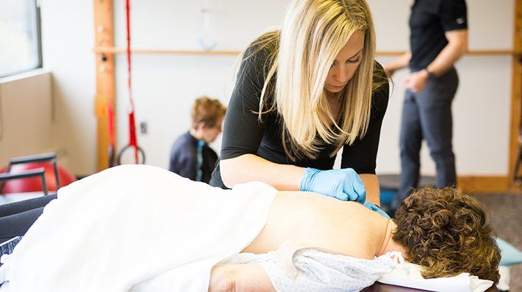
Dry needling is a technique physical therapists use (where allowed by state law) for the treatment of pain and movement impairments. The technique uses a "dry" needle, one without medication or injection, inserted through the skin into areas of the muscle. Other terms commonly used to describe dry needling, include trigger point dry needling, and intramuscular manual therapy. Dry needling is not acupuncture, a practice based on traditional Chinese medicine and performed by acupuncturists. Dry needling is a part of modern Western medicine principles and supported by research 1. What is a Trigger Point? A trigger point is a taut band of skeletal muscle located within a larger muscle group. Trigger points can be tender to the touch, and touching a trigger point may cause pain to other parts of the body. What Kind of Needles Are Used? Dry needling involves a thin filiform needle that penetrates the skin and stimulates underlying myofascial trigger points and muscular and connective tissues. The needle allows a physical therapist to target tissues that are not manually palpable. Physical therapists wear gloves and appropriate personal protective equipment (PPE) when dry needling, consistent with Standard Precautions, Guide to Infection Prevention for Outpatient Settings, and OSHA standards. The sterile needles are disposed of in a medical sharps collector. Why Dry Needling? In cases when physical therapists use dry needling, it is typically 1 technique that's part of a larger treatment plan. Physical therapists use dry needling to release or inactivate trigger points to relieve pain or improve range of motion. Preliminary research 2 supports that dry needling improves pain control, reduces muscle tension, and normalizes dysfunctions of the motor end plates, the sites at which nerve impulses are transmitted to muscles. This can help speed up the patient's return to active rehabilitation. As part of their entry-level education, physical therapists are well educated in anatomy and therapeutic treatment of the body. Physical therapists who perform dry needling supplement that knowledge by obtaining specific postgraduate education and training. When contacting a physical therapist for dry needling treatment, be sure to ask about their particular experience and education. www.choosept.com , the official consumer Website of the American Physical Therapy Association,© 2017
By Action PT June 24, 2020
Chronic pain is a condition that occurs when the brain concludes there is a threat to a person's well-being based on the many signals it receives from the body. This condition can and often does occur independently of any actual body tissue damage (due to injury or illness), and beyond normal tissue healing time. It is estimated that 116 million Americans have chronic pain each year. The cost in the United States is $560–$635 billion annually for medical treatment, lost work time, and lost wages. The causes of chronic pain vary widely. While any condition can lead to chronic pain, there are certain medical conditions more likely to cause chronic pain. These include: Trauma/injury Diabetes Mellitus Fibromyalgia Limb amputation Reflex Sympathetic Dystrophy Some diseases, such as cancer and arthritis, cause ongoing pain. With chronic pain, however, pain is created in the nervous system even after physical tissues have healed. Chronic pain affects each person experiencing it differently. In some cases, chronic pain can lead to decreased activity levels, job loss, or financial difficulties, as well as anxiety, depression, and disability. Physical therapists work together with chronic pain patients to lessen their pain and restore their activity to the highest possible levels. With treatment, the adverse effects of chronic pain can be reduced. For more resources on pain, and use of opioids for pain management, visit our Health Center on Pain and our Health Center on Opioid Use for Pain Management. The American Physical Therapy Association launched a national campaign to raise awareness about the risks of opioids and the safe alternative of physical therapy for long-term pain management. Learn more at our safe pain management page. What Is Chronic Pain? Pain is an unpleasant sensation that we usually associate with injury or tissue damage but can be present in the absence of tissue damage as well. Pain can be acute or chronic. Acute pain lasts for a short time – up to 12 weeks. It is a warning that tissue damage has occurred or may occur, or to help us prevent injury or disease. For instance, if we touch a hot stove, the body sends a danger message to the brain that there is a threat to tissues to prevent further injury. A sore foot can signal a need to change your footwear. In some cases, the danger messages may be due to some disease process, and your brain may interpret those messages as pain. This can cause you to seek medical attention – diagnosis and treatment – for what may be a serious condition. Signaling pain in this manner is the body's way of protecting us and is a good thing. Chronic pain is any discomfort or unpleasant sensation that lasts for more than three months – or beyond an expected normal healing time. Often, those who have chronic pain believe they have an ongoing disease or that their body has not healed, when this may not be the case. Chronic pain is likely not warning you of possible injury or danger; instead, the pain centers in the brain may be causing you to hurt even though there are no new causes of pain occurring in the body. Anyone can develop chronic pain at any age. The brain changes in chronic pain: When you are injured or develop a painful disease, nerves send information from the problem area to the brain. The brain analyzes this information coming from the body to determine if there is a threat to the body and whether action needs to be taken to prevent harm. When pain is constant or chronic, the brain and nervous system go on "high alert," becoming more sensitive. Cells that conduct sensation in the nervous system can also become more sensitive when on high alert, making it easier for the brain to interpret these sensations as a threat and thus cause you to have more pain. These changes in the brain and central nervous system induce and maintain chronic pain symptoms. When pain is chronic: Pain sensations are activated in the brain; The brain continues to interpret all sensations from the problem areas as a danger, even when there is no more tissue damage. This makes it easier for the pain centers in the brain to activate; Pain messages come from many different areas of the brain – areas that may control fight or fear reactions, movement, emotions, problem-solving, and learning. Almost any system of the body can be affected by chronic pain. The brain and nervous system continue reacting by causing you to continue to be in pain. This process increases sensations, emotions, or thoughts about the problem area. At this point, any sensory input can activate the pain centers. Even thinking about it or reading the word pain can trigger pain sensations. The pain is in the brain: To protect you, your brain is deciding to increase the alert level for sensations you feel. How Does It Feel? How chronic pain feels varies with each individual; it is very personal. How often it occurs, how severe it is, or how long it lasts is not predictable from one person to another. Common complaints related to chronic pain include: It may seem as if "everything hurts, everywhere." There may be sudden stabs of pain. It may seem as if the pain "has a mind of its own." You feel symptoms even if you are not doing anything to cause them. It feels worse when you think about it. It feels worse when you experience upsetting circumstances in your life. You may feel more anxious and depressed. You may feel your symptoms spread from one area to another area. You may feel fatigued, and afraid to do your normal activities. These complaints are common when you have chronic pain. However, it does not necessarily mean that your physical condition is worsening; it may just mean that your system has become more sensitive. Signs and Symptoms Research finds the following signs may be associated with a chronic pain syndrome: Fearfulness. It is easy to begin to fear increased pain when you have a chronic pain condition. As a result, you may begin to avoid activity. You may find that you rely more on family members to help with daily functions. Body stiffness when you try to become more active. Stiffness may make you feel as if your body is less able to perform daily activities. Deconditioning. Not moving your body results in less tolerance when you want to become more active. If you are inactive for a long time, muscles weaken and shrink from not being used. This can also increase your risk of falling. Decreased circulation. Lack of activity decreases the circulation of much-needed blood to your cells. Tissues in your body may not get as much oxygen as they need. As a result, they may not be as healthy as they can be. This can cause you to feel fatigued, and lack energy. Weight gain and/or a worsening of other conditions. Decreased activity can lead to unwanted weight gain. Added pounds and inactivity can aggravate symptoms of other conditions such as diabetes and high blood pressure. Chronic pain conditions are also commonly associated with feelings of anxiety or depression. Increased use of medication. Chronic pain patients can have the tendency to increase their medication over time to seek relief. Individual behaviors can include: Seeking out of many different doctors or health care providers and facilities to find relief. Difficulties with job performance. Some people with chronic pain even seek work disability. Avoidance of social situations or family members. When pain is ongoing, you may find you have feelings of bitterness, frustration, or depression. Some people report they have thoughts of suicide. If you are having these feelings, tell your doctor. This is important, so that you can get appropriate medications to help you feel better. How Is It Diagnosed? Your physical therapist will perform a thorough evaluation. He or she will: Ask specific questions about your past and present health and use of medication. Ask about your symptoms: their location, intensity, how and when the pain occurs, and other questions, to form a clear picture of your individual situation. Ask you to fill out pain and function questionnaires, to understand how the pain is affecting your daily life. Perform tests and movements with you. The tests help to identify problems with posture, flexibility, muscle strength, joint mobility, and movement. Special tests help to rule out any serious health problem such as pressure on a nerve or an underlying disease. Observe how you use your body for home, work, and social/leisure activities. This information helps your therapist prescribe a program that will boost your quality of life, and get you moving your best. Imaging tests such as x-rays, computed tomography (CT) scans, and magnetic resonance imaging (MRI) are often not helpful for diagnosing the cause of chronic pain. However, if your physical therapist suspects that your pain might be caused by any serious underlying condition, he or she will refer you to your physician for evaluation. Your physical therapist will work with your physician to provide the best diagnosis and treatment for your chronic pain. How Can a Physical Therapist Help? Your physical therapist will work with you to educate you on chronic pain, find solutions to improve your quality of life, and get you moving again! He or she will help you improve movement, teach you pain management strategies, and, in many cases, reduce your pain. Not all chronic pain is the same. Your therapist will evaluate your clinical examination and test results and design an individualized treatment plan that fits you best. Physical therapy treatments may include: Education to improve your knowledge and understanding of chronic pain -- how it occurs and what you can do about it. Your therapist will teach you how to manage your pain and help you work toward performing your normal daily activities again. Strengthening and flexibility exercises to help you move more efficiently with less discomfort. Your therapist will design a program of graded exercises for you -- movements that are gradually increased according to your abilities. Graded exercises help you improve your coordination and movement, reducing the stress and strain on your body and decreasing your pain. Carefully introducing a graded exercise program will help train your brain to sense the problem area in your body without increasing its danger messages. Manual therapy, which consists of specific, gentle, hands-on techniques that may be used to manipulate or mobilize tight joint structures and soft tissues. Manual therapy is used to increase movement (range of motion), improve the quality of the tissues, and reduce pain. Posture awareness and body mechanics instruction to help improve your posture and movement. This training enables you to use your body more efficiently while performing activities and even when you are resting. Your therapist will help you adjust your movement at work, or when performing chores or recreational activities, to reduce your pain and increase your ability to function. The use of ice, heat, or electrical stimulation has not been found to be helpful with chronic pain. Your physical therapist, however, will determine if any of these treatments could benefit your unique condition. www.choosept.com , the official consumer Website of the American Physical Therapy Association,© 2017
By Action PT June 24, 2020
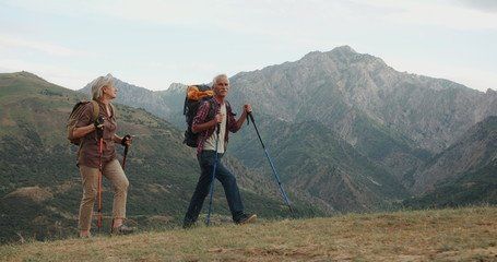
Aging can have a bad reputation; however, there is no reason that growing older can’t be a journey full of adventure and opportunities to live life to the fullest. All you need is proper guidance, proactive steps, and a positive attitude. And there is proof. Research suggests that improvements in physical function are possible well into older adulthood, and supports that continued activity as you age helps fight cognitive decline. With the guidance of a physical therapist, you can improve mobility, maintain your independence, and continue participating in your favorite daily activities. As a movement expert, a physical therapist can provide an evaluation and design a treatment program to address any of your ailments and deficiencies, make modifications based on other preexisting conditions, and help you achieve your goals. The following tips can keep you active and independent as you age: Staying fit. Whether you are participate in regular exercise or just want to stay fit for daily activities, exercise is necessary. Exercise is proven to help improve balance, strengthen bones, and prevent heart and brain conditions. A prescribed strength training and aerobic exercise program will help you maintain and strengthen critical muscle groups needed for your life. Staying balanced. Maintaining balance and avoiding falls are imperative to maintaining a quality of life and living independently. A physical therapist can prescribe a customized program of static and dynamic balance activities and exercises to improve your balance and prevent dangerous falls. Assessing the terrain. A physical therapist can make recommendations that make your home and other environments safe by eliminating dangerous barriers. Typically this means removing throw rugs, loose carpets, clutter, and modifying entry thresholds with ramps. Staying engaged. Successful aging is not only impacted by one’s environment, but also how one interacts with their environment. Fear and social isolation are substantial factors that increase fall risk and jeopardize independence. To ensure active and successful aging, continue to engage with friends, family, and the community. Social experiences with friends and family help diminish fear and improve physical and mental capabilities. www.choosept.com , the official consumer Website of the American Physical Therapy Association,© 2017
By Action PT June 24, 2020
Stephanie Carter Kelley, PT, PhD, discusses how she’s incorporated yoga into her physical therapist practice. This video was produced by the Ohio Physical Therapy Association. https://youtu.be/z43LmxvRbSU www.choosept.com , the official consumer Website of the American Physical Therapy Association,© 2017
By Action PT June 24, 2020
Physical therapist Sara Bellanca, PT, DPT, leads fun physical activities for kids using popsicle sticks drawn from a cup to prompt each new movement activity. https://youtu.be/pN19I9XPtRA Sara received a doctor of physical therapy degree from Ithaca College. She is a school-based physical therapist who works with children who have developmental disabilities at Mary Cariola Center in Rochester, New York. Her passion lies in helping children of all abilities have fun doing physical activities, as they work on important skills that are crucial to their development, function, and independence. www.choosept.com , the official consumer Website of the American Physical Therapy Association,© 2017
By Action PT June 24, 2020
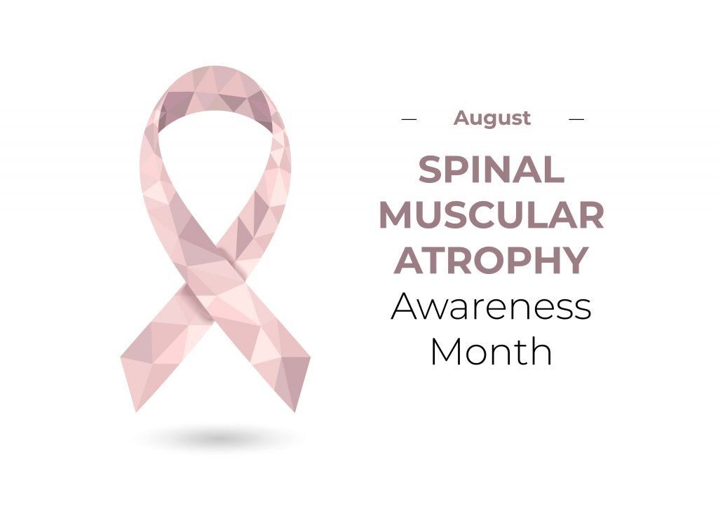
Spinal muscular atrophy (SMA) is a common, inherited neuromuscular disease that causes low muscle tone (hypotonia) and progressive muscle weakness and wasting (atrophy). All motor skills can be affected by the disease, including walking, eating, and breathing. SMA is a leading cause of death in infants. It affects approximately 1 in 10,000 babies born in the United States, of any race or gender. Approximately 1 in every 50 Americans is a genetic carrier of SMA. Physical therapists help children with SMA develop muscle strength and movement abilities to function at the highest level possible. What is Spinal Muscular Atrophy? Spinal muscular atrophy (SMA) is a genetic (inherited) neuromuscular disease. The large motor nerves of the spinal cord are abnormal in people with SMA because a gene (the Survival Motor Neuron-1 or SMN1 gene) is missing or altered. Without the gene, or with a damaged gene, the nerves do not have a specific protein that allows them to control muscles. The decrease of the SMN1 protein results in improper functioning, and eventually death of the motor nerve cells in the spinal cord. The severity of SMA is related to the amount of the SMN1 protein that is absent in the motor nerve cells (more protein allows for more function). Severity of the disease ranges from mild muscle weakness, to total paralysis and the need for support to breathe. Signs and Symptoms Muscle weakness is a hallmark feature of SMA. The muscle weakness is associated with other impairments, such as: Muscle wasting (atrophy). Low muscle tone that makes the child’s body feel loose or “floppy” (hypotonia). Absent or markedly decreased deep tendon reflexes (eg, knee-jerk reflex). Involuntary tongue movements. Hand tremors. Tight joints that do not bend or stretch all the way, or possibly don’t move at all. Spinal deformity (scoliosis). Respiratory infections and lung disease. The course of SMA varies with each individual, and symptoms range from mild to severe. The onset of SMA can occur from birth to adulthood. The progression of the disease varies, but generally as an individual ages, muscle strength decreases and symptoms progress. However, some people with SMA live a normal lifespan. How Is It Diagnosed? Diagnosis of SMA begins by observing whether an individual has any of the signs and symptoms of the disease. When SMA is suspected, a simple blood test can identify an estimated 95% of all cases of SMA by revealing the specific mutation that causes the disease. The other 5% of cases are caused by a rare mutation, and must be identified through further testing. A muscle biopsy may be performed to identify any nerve deterioration, and electromyography (EMG) will test how well the nerves are controlling the muscles. When diagnosed, SMA is classified into 1 of several types: Type I SMA (Werdnig–Hoffmann Disease). Type I SMA is the most frequently diagnosed and most severe form of SMA. The disease is apparent at birth or in the first 6 months of life, and the child never learns rolling or independent sitting. The child has severe, generalized muscle weakness, and low muscle tone (hypotonia). Infants typically have muscle wasting (atrophy), weakness in the head and neck, arms, legs, and trunk, and decreased movement. Involuntary movements of the tongue and tremors in the hand may be seen. The child may develop respiratory infections. Type II SMA (Intermediate Form). In Type II SMA, the disease develops between the ages of 6 months and 18 months, and causes less severe weakness than Type I SMA. Children with Type II SMA are able to learn to sit up and to stand, but usually are not able to walk independently without braces and assistive devices, such as walkers or crutches. Tongue movements and hand tremors may be seen, but are less apparent than in Type I SMA. Contractures (tightening) of joints and scoliosis are common. Respiratory infections and complications occur as the disease progresses, and the need for help with breathing is common in the later stages of the disease. Type III SMA (Kugelberg–Welander Disease). Type III SMA has a later onset and better prognosis. Type III SMA is usually diagnosed between the ages of 18 months and 3 years, or even later in mild cases, where weakness is not noticeable until late childhood. Children are able to achieve developmental milestones and independent walking, and often they maintain walking until adolescence or adulthood. Walking may be characterized by a lack of balance, frequent falls, scoliosis, knee extension that is more than usual and causes the knee to “bend backward” slightly, and excessive waddling. The muscle weakness mainly affects the muscles around the hips, and is less severe than in Types I and II. Rare SMAs Other forms of SMA are rare and are caused by different genes than the SMN1 gene. These forms of SMA include distal spinal muscular atrophy, Kennedy disease, and other classifications. Your physical therapist will educate you about the rarer forms of the disease. How Can a Physical Therapist Help? Evaluation is important for guiding the treatment of a child with SMA. Your physical therapist will conduct a thorough evaluation that includes taking the child’s health history and examining the child’s posture, muscle strength, motor activities that include walking, joint motion, respiratory function, participation with family and friends, and quality of life. Treatment is important to allow children with SMA to achieve the highest level of independent living and mobility possible, and to prevent or delay the development of complications. Physical therapists work closely with the child and family to develop the most appropriate goals for each child based on functional levels and interests. Your child's treatment may include: Therapeutic Exercises and Strength Training. Physical therapists use different therapeutic exercises to help children with SMA improve and maintain mobility, and prevent or slow the progression of contractures and respiratory failure. Therapeutic exercises may include strengthening and aerobic at levels appropriate for the specific child. Strengthening Exercises. Your physical therapist can help your child slow any deterioration in muscle strength and prolong your child’s ability to walk. Physical therapists design strengthening exercises to keep children with SMA active and moving. They identify games and fun tasks that can be used during therapy or taught to the family to maintain strength. Improving Developmental Skills. Your physical therapist will develop strategies to help your child learn developmental skills that will improve: Head and trunk control Floor mobility, such as rolling and crawling Changing positions, such as pulling up to stand Learning upright positions and skills, such as sitting, standing, and walking Aquatic Therapy or Hydrotherapy. Some physical therapists specialize in aquatic therapy and use the physical properties of water to provide strength training, walking and balance exercises, and aerobic training, without the risk of fatigue or overworked muscles. Standing Programs. Standing programs for children who can't walk are used to maintain muscle flexibility and length, prevent contractures, promote musculoskeletal development, and prevent bone-mineral density loss. Your physical therapist can design standing programs to be used at home or at school, as appropriate. Management of Respiratory Complications. Physical therapists teach parents and caregivers how to perform chest drainage techniques, and help with coughing and breathing techniques to keep children with SMA breathing well. Your physical therapist will provide chest physical therapy as appropriate, and teach the family some blowing games (like bubbles) to improve breathing. Management of Contractures. Physical therapists help prevent joint tightness (contractures) and increase flexibility in children with SMA by designing specific programs in range-of-motion exercises, positioning, and regular stretching. Your physical therapist may recommend the use of splints, braces, or standing devices, as appropriate. Feeding. Physical therapists work closely with speech or occupational therapists to promote healthy feeding in children with SMA. They will help position the child in appropriate head and body postures to allow the most effective feeding. Management of Scoliosis and Skeletal Deformities. Physical therapists can assist in the prevention or reduction of scoliosis and skeletal deformities by designing specific programs to improve movement, and maintain healthy positioning at all times. They will recommend wheelchair modifications and the use of braces to ensure the safety and health of each individual child, as needed. If a child requires surgery for scoliosis or other joint deformities, intensive preoperative and postoperative physical therapy can help prevent respiratory complications and loss of strength or function. Assistive Devices. Many children with SMA require adaptive or assistive devices to help them maintain function at some point during the course of the disease, and especially following surgery. Physical therapists work with other rehabilitation specialists to select and modify assistive devices to meet each individual child's specific needs. Types of assistive devices range from those that position a child for feeding or playing, to motorized wheelchairs. Can This Injury or Condition Be Prevented? SMA is a genetic disorder that is passed from parent to child. Most people have 2 copies of the SMN1 gene. Individuals who have 1 faulty copy and 1 functioning copy of the gene are called carriers. Carriers do not have SMA, but they may pass the faulty gene on to their children. SMA is an autosomal recessive genetic disorder, which means that generally both parents must pass on the mutation for the child to have SMA. Because the specific mutation that causes SMA has been identified, genetic testing using a simple blood test can identify carriers of SMA. However, most people do not know they are carriers until they have a child with SMA. Excellent prenatal care is important for all pregnant women. Testing can be performed during the pregnancy to determine if a baby has SMA. Once a child is diagnosed with SMA, physical therapists and other health care professionals can reduce some of the additional complications that occur following birth, such as developmental delay, poor feeding, abnormal postures and scoliosis, loss of joint range of motion, abnormal movement or walking patterns, and respiratory problems. www.choosept.com , the official consumer Website of the American Physical Therapy Association,© 2017
By Action PT June 24, 2020
De Quervain's (dih-kwer-VAINS) tendinitis is a condition that causes pain and tenderness at the thumb side of the wrist, at the base of the thumb and forearm. Pain is worsened with grasping or extending the thumb (pulling it back like "thumbing a ride"). People of all ages can develop this condition, which usually happens when the tendons are strained by prolonged or repetitive use of the hand, rapid or forceful hand use, or use of the hand or arm in an awkward position. Tendons at the wrist become irritated and thickened, resulting in pain when moving the thumb and grasping objects. Common forms of treatment for De Quervain’s include splinting and range-of-motion exercises. Injection for cortisone by a doctor is common treatment. Persistent cases may require surgery. What is De Quervain’s Tendinitis? De Quervain's tendinitis is a common condition involving tendons of the wrist. Tendons are tough cords or bands of connective tissue that attach muscles to a bone. The thumb and wrist extensor tendons (Abductor Pollicus Longus and Extensor Pollicus Brevis) are encased in a "sheath" or a tunnel at the wrist, which keeps the tendons in place. De Quervain's tendinitis can occur gradually or suddenly, when the tendons become inflamed or thickened from overload or repetitive use, and have difficulty sliding through the extensor tunnel. Risk factors for developing De Quervain's tendinitis include: Chronic overuse of the hand. Excessive use of the thumb from texting and gaming. Being female (women are 8 to 10 times more likely to develop this condition than men). Pregnancy. Using the hand or arm in a position that feels awkward. Participation in sports that stress the hand and wrist, such as golf and tennis. Age greater than 40 years. Race; members of the black community may be more likely to experience this problem. How Does It Feel? A person who has De Quervain's tendinitis may: Feel localized tenderness, pain, and, swelling at the wrist near where the thumb is attached to the forearm. Have difficulty pinching or grasping with the thumb or hand. Feel pain when moving the wrist from side to side or twisting it. Experience limited motion and feeling of weakness in the thumb. Have difficulty flexing the thumb. Notice a "catching" or "snapping" sensation with movement of the thumb (a less common symptom). Active use of the hand with activities, such as writing, opening jars, lifting a child, hammering, sports, and any workplace or home activity that involves pinching or grasping with the thumb, can provoke the symptoms of pain, stiffness, and weakness. Note: Other conditions of the wrist and hand can cause symptoms similar to those stated here. Your physical therapist will help to identify any underlying problems of your joints, tissues, or nerves that may be causing similar symptoms. How Is It Diagnosed? Your physical therapist will ask you how and when you first experienced symptoms, and what it feels like at the present time. Your therapist will perform a physical exam that will include feeling for tender spots, measuring the flexibility and range of motion of the thumb and wrist, and testing the strength of the thumb muscles and grip. Your physical therapist will also perform a Finkelstein test, which gently stretches the tendons on the thumb side of the wrist through the extensor tunnel. Pain during this test is common with De Quervain’s tendinitis. Your physical therapist will also perform other tests to rule out any underlying conditions that may mimic De Quervain's symptoms. How Can a Physical Therapist Help? If your physical therapist confirms De Quervain’s Tendinitis from an evaluation, they will work with you to develop an individualized plan with you for this condition. Your physical therapist will review and evaluate how you use your hand and wrist for functional activity. The review will include your daily activities, work, and sports activities. The physical therapist will try to help you identify what activities or positions that you use that may contribute to the problem. They will instruct you how to make changes in your function to help healing and reduce risk of the problem in the future. Specific instructions may include avoiding repetitive thumb and/or wrist movements, avoid flexing the thumb, and avoid moving the hand toward the pinkie finger as much as possible. You should also avoid forceful hand movements, and any movements or activities that increase pain. The therapist may provide a wrist splint to position your wrist and thumb for rest, and to provide compression to help pain and swelling. Your therapist may also work with you to reduce pain and inflammation. Ultrasound therapy may be applied to improve pain. This treatment uses ultrasonic sound waves applied over the involved area to improve circulation, reduce swelling, and aide healing of the tissues and tendons. Iontophoresis is another option to reduce swelling and pain. Iontophoresis is a type of electrical stimulation that is used to administer medication to the problem area through your skin. Ice or heat may be recommended for short term pain relief. Your therapist will advise you for what is best for your condition. Exercise is prescribed to improve range of motion and prevent stiffness. Early on, exercise is restricted to avoid aggravating the condition. As the condition improves, exercises will be progressed to improve strength for functional activity, as well as improve active range of motion of the thumb and wrist. If your symptoms do not respond to conservative care, your physical therapist will refer you to a physician who will determine if you need medication, injection, or surgical care for further recovery. How Can a Physical Therapist Help Before & After Surgery? If your De Quervain's problem does require surgery, your physical therapist may fit a splint to your hand and wrist after the procedure. Your physical therapist will help you to control any swelling, maintain and improve your hand and wrist flexibility, build your strength, and improve your range of motion, allowing you to safely return to your preinjury activity levels. Can This Injury or Condition Be Prevented? It may be possible to prevent De Quervain's tendinitis. Some risk factors cannot be controlled, such as gender, race, or age; however, physical therapists recommend that you: Avoid chronic overuse of the hand. Avoid or restrict overly forceful use of the wrist. Avoid excessive use of the thumbs for texting and gaming. Avoid putting the wrist and hand in awkward positions while using the hand or arm. Train and condition in sports, such as golf and tennis to minimize wrist and thumb strain. Your physical therapist can teach you correct and safe hand and wrist positions to maintain during your daily home, work, and sport activities. www.choosept.com , the official consumer Website of the American Physical Therapy Association,© 2017
By Action PT June 24, 2020
Common gardening activities, such as digging, planting, weeding, mulching, and raking can cause stress and strain on muscles and joints. The following tips can help minimize aches and pains: 1. Get moving before you garden. A 10 minute brisk walk and stretches for the spine and limbs are good ways to warm up. 2. Change positions frequently to avoid stiffness or cramping. Be aware of how your body feels as you work in your garden. If a part of your body starts to ache, take a break, stretch that body part in the opposite direction it was in, or switch to a different gardening activity. For example, if you've been leaning forward for more than a few minutes, and your back starts to ache, slowly stand up, and gently lean backwards a few times. 3. Use a garden cart or wheelbarrow to move heavy planting materials or tools. Lift with your knees and use good posture while moving a cart or wheelbarrow. 4. Give your knees a break. Use knee pads or a gardening pad. If kneeling or leaning down to the ground causes significant pain in your back or knees, consider using elevated planters to do your gardening. If kneeling on both knees causes discomfort in your back, try kneeling on one and keep the other foot on the ground. 5. Maintain good posture. Use good body mechanics when you pick something up or pull on something, such as a weed. Bend your knees, tighten your abdominals, and keep your back straight as you lift or pull things. Avoid twisting your spine or knees when moving things to the side; instead, move your feet or pivot on your toes to turn your full body as one unit. 6. Take breaks. If you haven't done gardening or other yard work in a while, plan to work in short stints, building in time for breaks before you start feeling aches and pains. 7. Keep moving after you garden. End your gardening session with some gentle backward bending of your low back, a short walk and light stretching, similar to stretches done before starting. If your aches and pains aren't managed with these tips and discomfort lingers, a physical therapist can help. www.choosept.com , the official consumer Website of the American Physical Therapy Association,© 2017
By Action PT June 24, 2020
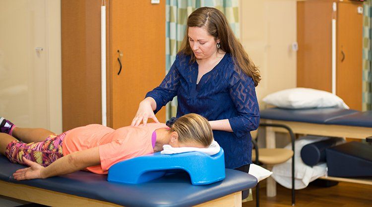
Physical therapy is among the safe and effective alternatives to opioids recommended by the Centers for Disease Control and Prevention for the management of most non-cancer related pain. Whereas opioids only mask the sensation of pain, physical therapists treat pain through movement, hands-on care, exercise, and patient education. Here's how physical therapists manage pain: 1. Exercise. A study following 20,000 people over 11 years found that those who exercised on a regular basis, experienced less pain. And among those who exercised more than 3 times per week, chronic widespread pain was 28% less common1. Physical therapists can prescribe exercise specific to your goals and needs. 2. Manual Therapy. Research supports a hands-on approach to treating pain. From carpal tunnel syndrome2 to low back pain3, this type of care can effectively reduce your pain and improve your movement. Physical therapists may use manipulation, joint and soft tissue mobilizations, and dry needling, as well as other strategies in your care. 3. Education. A large study conducted with military personnel4 demonstrated that those with back pain who received a 45 minute educational session about pain, were less likely to seek treatment than their peers who didn't receive education about pain. Physical therapists will talk with you to make sure they understand your pain history, and help set realistic expectations about your treatment. 4. Teamwork. Recent studies have shown that developing a positive relationship with your physical therapist and being an active participant in your own recovery can impact your success. This is likely because physical therapists are able to directly work with you and assess how your pain responds to treatment. Read more about Pain and Chronic Pain Syndromes. The American Physical Therapy Association launched a national campaign to raise awareness about the risks of opioids and the safe alternative of physical therapy for long-term pain management. Learn more at our #ChoosePT page. www.choosept.com , the official consumer Website of the American Physical Therapy Association,© 2017
By 06/22/20 June 22, 2020
Stroke (when a blood vessel in the brain is blocked or ruptured) is the third leading cause of death in the United States, and is a leading cause of serious, long-term disability in adults. Stroke can happen to anyone at any time—regardless of race, sex, or even age—but more women than men have a stroke each year, and African Americans have almost twice the risk of first-ever stroke than Caucasians. Approximately two-thirds of those who experience a stroke are over 65 years of age. Almost 800,000 people in the United States have a stroke each year. Physical therapists provide treatments for people who have experienced stroke to restore their movement and walking ability, decrease their disability, and improve their quality of life. If you have 1 or more of the following symptoms, immediately call 911 or emergency medical services (EMS) so that an ambulance can be sent for you: Sudden numbness or weakness of the face, arm, or leg, especially on one side of the body Sudden confusion or trouble speaking or understanding Sudden trouble seeing in one or both eyes Sudden trouble walking, dizziness, loss of balance or coordination Sudden, severe headache with no known cause If You Think Someone Might Be Having a Stroke Act F.A.S.T.! Emergency treatment with a clot-buster drug called t-PA can help reduce or even eliminate problems from stroke, but it must be given within 3 hours of when symptoms start. Recognizing the symptoms can be easy by remembering to think F.A.S.T. F=Face. Ask the person to smile. Does one side of the face droop? A=Arms. Ask the person to raise both arms. Does one arm drift downward? S=Speech. Ask the person to repeat a simple phrase. Does the speech sound slurred or strange? T=Time. If you observe any of these signs, call 911 and note the time that you think the stroke began. Research shows that people with stroke who arrive at the hospital by ambulance receive quicker treatment than those who arrive by their own means. What Is Stroke? Stroke is a term used when a blood vessel in the brain is blocked (65% of all strokes) or ruptures. It is also called a cerebral vascular accident (CVA). If the blood flow is stopped or altered, a part of the brain does not receive enough oxygen. Millions of brain cells die every minute during a stroke, increasing the risk of permanent brain damage, disability, or death. An ischemic stroke, the most common type, occurs when a blood vessel is blocked. One frequent cause of blockage is a blood clot or a build-up of fatty deposits (arteriosclerosis) in blood vessels that supply the brain. A hemorrhagic stroke occurs when a blood vessel leaks or ruptures because of a weakened region of a vessel or in an area of the brain with a cluster of abnormally formed vessels. Signs and Symptoms If you are having a stroke, you might: Feel a sudden numbness or weakness of the face, arm, or leg, especially on one side of the body Be confused about where you are or what you're doing Have trouble speaking or understanding what others are saying Have trouble seeing in one or both of your eyes Have trouble walking, be dizzy, or lose your balance Have a sudden, severe headache that seems to come out of nowhere Some people experience a transient ischemic attack (TIA), a "mini-stroke" that produces stroke-like symptoms, but no permanent damage because the blood flow is altered for only a short period of time. Recognizing and seeking treatment for TIAs can reduce your risk of a major stroke. Stroke can cause a range of long-term problems, such as: Inability or difficulty moving one side of the body (hemiparesis or hemiplegia) Severely limited movement or stiffness in the arms and legs (spasticity) Balance problems Weakness on one side of the body Off-and-on numbness Loss or lack of sensation Sensitivity to cold temperatures Memory loss Slowed or slurred speech Difficulty remembering words How Is It Diagnosed? Physicians or emergency medical personnel will assess a stroke based on the signs and symptoms. Stroke is often confirmed by examining the patient, using clinical tests and taking images of the brain, usually with a CT scan or MRI. Since research has proven that early treatment can reduce the effects of stroke and save lives, efforts are being made to get persons suspected of having a stroke to a hospital stroke unit, rather than the emergency department. Physicians may use a classification system, called TOAST, to determine the type of stroke, so that treatment can begin quickly. How Can a Physical Therapist Help? Physical therapists are part of the stroke rehabilitation team. Rehabilitation begins very soon after a stroke; your physical therapist’s main goal is to help you return to your activities at home, at work, and in your community. After examining you and evaluating your condition, your physical therapist will develop an individualized plan to help you achieve the best possible quality of life. The plan will focus on your ability to move, any pain you might have, and ways to prevent problems that may occur after a stroke. One of the first things your physical therapist will teach you is how to move safely from your bed to a chair, and to perform exercises in bed. As you become more mobile, your physical therapist will teach you strengthening exercises and functional activities. Later, your physical therapist will: Help you improve your balance and walking. Fit you with a wheelchair, if needed. Provide training to your family and caregivers. Train you in how to use devices that can help you keep mobile when a stroke has affected your ability to move, walk, or keep your balance: orthoses, prostheses, canes, walkers, wheelchairs, and perhaps even robotics. Treatment for stroke varies. Your specific treatment will depend on the results of your physical therapist's evaluation, and on how long it's been since you had your stroke. Recovery from a stroke depends on the size and location of the stroke, how quickly you received care, and your other health conditions. Your physical therapist will help you regain functional skills to allow you to participate in your specific life activities. Relearning How to Use Your Upper Body, How to Walk, and How to Perform Daily Activities Your physical therapist will design an exercise and strengthening program based on tasks that you need to do every day, selecting from a variety of treatments. Physical therapist researchers are at the forefront of innovating many of these techniques: Constraint-induced movement therapy (CIMT). CIMT is used to strengthen an arm affected by the stroke. Your physical therapist will apply a mitten or a sling on your strong arm to keep you from fully using it. This constraint "forces" you to use your arm or hand affected by the stroke to perform daily tasks, which helps build your strength and control. CIMT requires that the constraint be used for several hours a day, several days a week to be successful. Functional electrical stimulation (FES). This treatment helps move your muscles if they are very weak. For instance, your physical therapist might use FES to treat a painful or stiff shoulder. Motor imagery and mental practice. These tools are used to help strengthen the arms, hands, feet, and legs. Working with your physical therapist, you will "rehearse" a movement without actually performing it, which stimulates the part of your brain that controls the movement. Positioning. Proper positioning helps reduce any muscle pain, spasms, slowness, or stiffness that can result from stroke. Your physical therapist will teach you how to safely move ("transfer") from a sitting to a standing position, and how to support yourself when sitting or lying down, using foam wedges, slings, and other aids. Virtual reality and interactive video games may be used to provide experiences similar to real life. Using a keyboard and mouse, a special wired glove, or sensors on your body, you can practice daily tasks as your therapist helps you "rewire" your brain and nerve connections. Your therapist may teach you how to continue these activities at home. Partial body weight support (BWS). BWS is used to help support you as you walk, usually on a treadmill. Your physical therapist will gradually decrease the amount of support as your posture, strength, balance, and coordination improve. Biofeedback. This treatment helps make you aware of how your muscles work and how you might be able to have better control over them. Your therapist will attach electrodes to your skin to provide measurements of muscle activity that are displayed on a monitor. Your physical therapist will work with you to help you understand and change those readings. Your needs will change over time, and your physical therapist may consider using aquatic therapy, robotics, or support devices to assist in your recovery. Even after rehabilitation is completed in a facility, your physical therapist will continue to see you as needed to assess your progress, update your exercise program, help you prevent further problems, and promote the healthiest possible lifestyle. . www.choosept.com , the official consumer Website of the American Physical Therapy Association,© 2017
Show More
Share by: